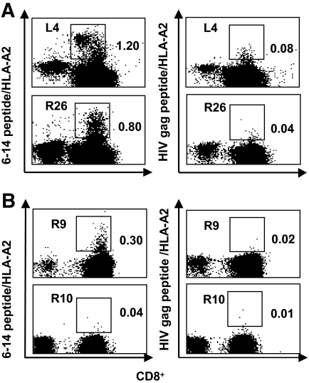
FIG. 3.
Peptide/HLA-A2 tetramers staining of CD8+ T-cells from diabetic patients. A: Staining of expanded CD8+ T-cells from two diabetic patients. PBMCs from one long-standing (L4) diabetic patient and one recent-onset (R26) diabetic patient were cultured in presence of peptides (peptide 6–14 or HIV gag peptide) and then stained by 6–14 peptide/HLA-A2 and HIV gag peptide/HLA-A2 tetramers. Cultures were harvested for tetramer staining on day 14. HIV gag peptide/HLA-A2 tetramer was used as a negative control. Detection of tetramer-stained cells was performed by gating out 7-AAD+ dead cells and selecting CD8+ T-cells. The figure shows the staining with 6–14 peptide/HLA-A2 tetramer (left) and with HIV gag peptide/HLA-A2 tetramer (right) at day 14 of cell expansion. Dot plots show tetramer (vertical axis) versus CD8+ cells (horizontal axis) staining. The numbers displayed in each dot plot indicate the percentages of cells stained by tetramers. B: Ex vivo tetramer staining of PBMCs from 6–14 peptide ELISpot-positive and ELISpot-negative diabetic patients. PBMCs from 6–14 peptide ELISpot-positive diabetic patients were stained ex vivo with 6–14 peptide/HLA-A2 and HIV gag peptide/HLA-A2 tetramers. The figure shows the ex vivo staining with 6–14 peptide/HLA-A2 tetramer (left) and with HIV gag peptide/HLA-A2 tetramer (right) for one 6–14 peptide ELISpot-positive patient (R9). As a control, a 6–14 peptide ELISpot-negative patient (R10).

