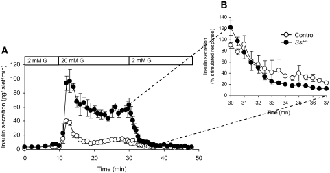
FIG. 5.
Rate of decline in insulin secretion from control and Sst−/− islets after removal of glucose stimulus. A: Glucose-induced (20 mmol/l G, bar) insulin secretion from Sst−/− islets and control islets is reversed to basal levels upon removal of stimulus. B: The decline in insulin secretion from stimulated to basal levels is expressed as percent stimulated insulin secretion before removal of glucose. Points show means ± SE, n = 4 separate perifusion channels.

