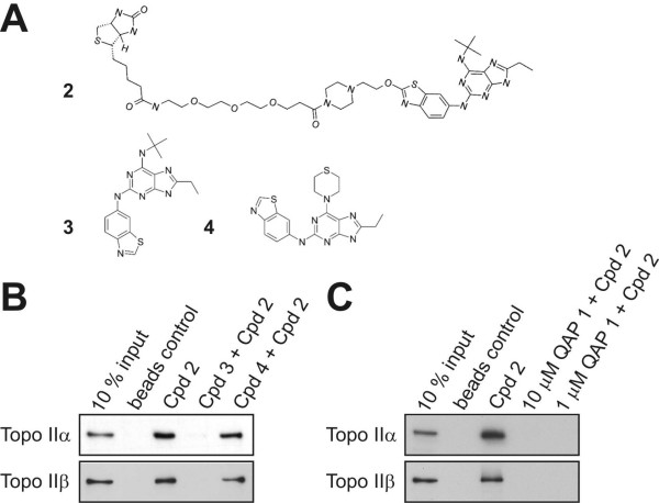
Figure 2.
Cell-free topoisomerase II alpha and beta interaction studies with substituted purine analogues. (A) Chemical structures of biotinylated purine analogue 2 (compound (cpd) 2), purine analogues 3 (cpd 3) and 4 (cpd 4), respectively. (B) Binding of both topoisomerase II alpha and beta to 1 μM biotinylated purine analogue 1 and disruption of binding by parent compound 3 but not ATPase inactive compound 4 (both at 10 μM). (C) Binding of topoisomerase II alpha and beta to 1 μM biotinylated compound 1 is abolished by 10 fold molar excess and equimolar concentrations of QAP 1.

