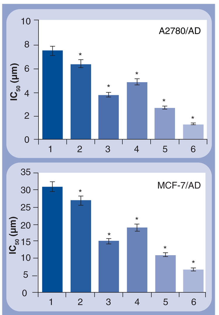
Figure 8. Cytotoxicity of different formulations in human multidrug-resistant ovarian (A2780/AD) and breast (MCF-7/AD) cancer cells.
The cells were incubated for 48 h with the indicated formulations. The concentration of siRNA and composition of cationic liposomes in all formulations were the same. Means ± standard deviation are shown. 1: Free DOX; 2: Cationic liposomes–DOX; 3: Cationic liposomes–BCL2 siRNA; 4: Cationic liposomes–DOX–MDR1 siRNA; 5: Cationic liposomes-MDR1 siRNA–BCL2 siRNA + Free DOX; 6: Cationic liposomes–DOX–MDR1 siRNA–BCL2 siRNA.
*p <0.05 when compared with free DOX.

