
Figure 2.
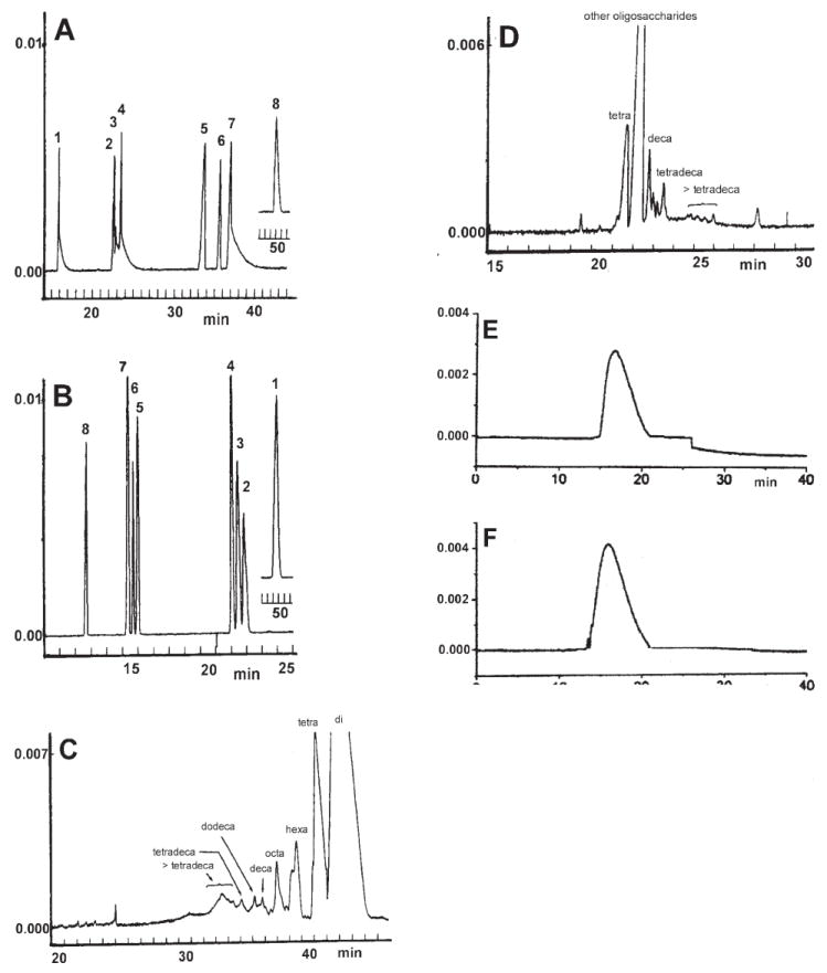
Electropherograms of (A) eight heparin-disaccharides 1–8 (1 is unsulfated and 8 is trisulfated, see ref. [76] for structures) using normal polarity in 10 mM sodium borate buffer and 50 mM SDS (SB-SDSB), pH 8.8, 12 kV. (B) Eight Hep-disaccharides 1–8 using reversed polarity in 20 mM sodium phosphate buffer pH 3.48 (SPB) at 12 kV. (C) Hep-oligosaccharides prepared by partial Hep lyase I digest (30% complete) of Hep using normal polarity at 12 kV. (D) Hep-oligosaccharides prepared by partial Hep lyase I digest (30% complete) of Hep using reversed polarity at 12 kV. (E) Hep using reversed polarity at 20 kV. (F) Low-molecular-weight Hep (MW 4800, RD heparin, Wyeth) using reversed polarity at 20 kV. (A) and (C) were in 10 mM sodium borate buffer and 50 mM SDS, (B) and (D) were in 20 mM sodium phosphate buffer pH 3.48, (E) and (F) were in 5 mM copper(II) sulfate. (A–D) were detected at 232 nm [76] and (E) and (F) were detected at 240 nm [55] (modified from ref. [76, 55]).