
Figure 3.
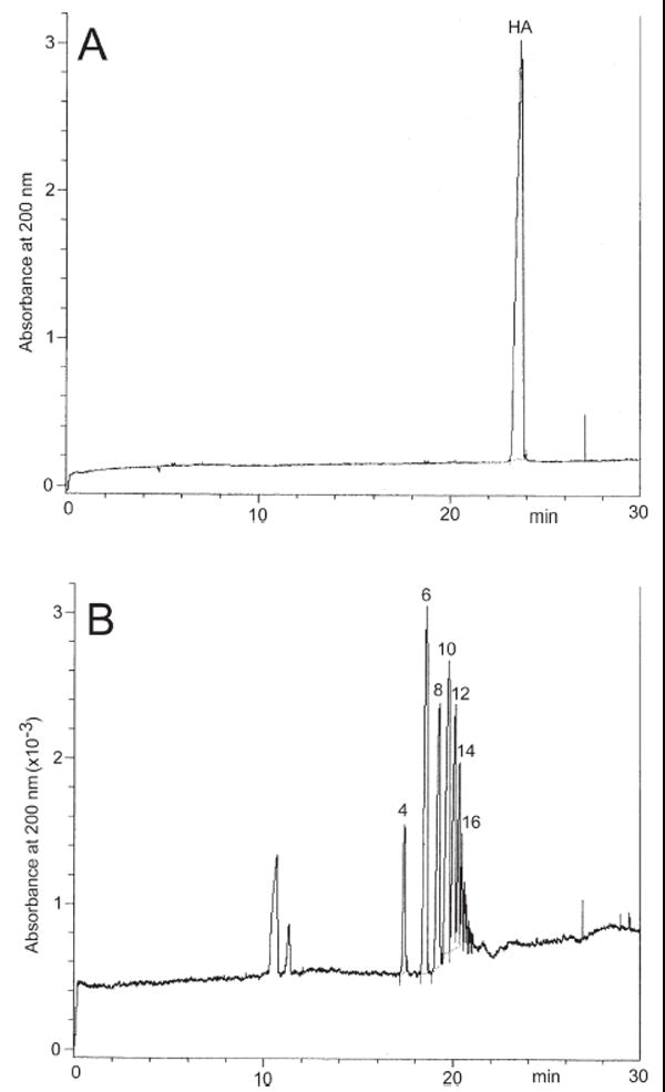
Electropherograms of (A) intact HA and (B) HA treated with hyaluronidase. CE was performed on a Beckman HPCE instrument (P/ACE system 5000) equipped with a UV detector set at 200 nm. Separation and analysis by EKC with SDS were carried out on an uncoated fused-silica capillary tube (50 μm id, 85 cm total length and 65 cm from the injection point to the detector) at 25°C. The operating buffer was constituted of disodium hydrogen phosphate (40 mM), sodium tetraborate (10 mM), and SDS (40 mM) buffered at pH 9.0. HA oligomers given above the peaks.