Fig. 3.
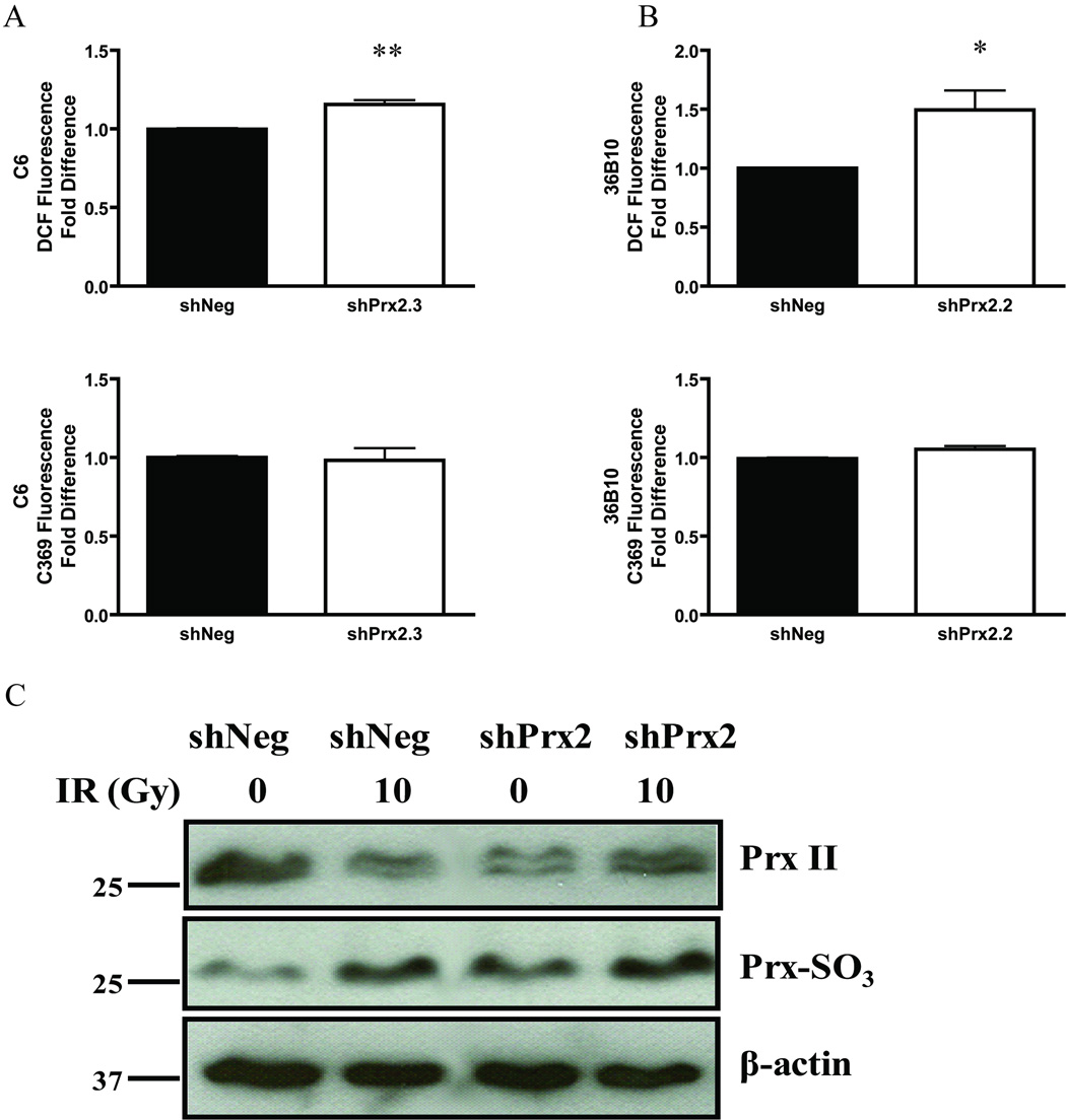
Exogenous H2O2 increases intracellular ROS generation. Cells were treated with 100 µM H2O2 and intracellular ROS generation was examined by DCF fluorescence in C6 (A) and 36B10 (B) cells; increases were observed in the shPrx2 cell lines. No alterations were detected using the non-oxidizable probe (A–B) for C6 and 36B10 cell lines, respectively. Western blot analysis was used to detect inactive hyperoxidized Prxs. Cells were either sham irradiated or irradiated with 10 Gy and probed for protein expression 24 h following IR. Immunoblots show an elevation in hyperoxidized Prxs in shPrx2 cells when compared to shNeg controls and levels were furthered elevated following IR (C). Representative blot of two independent experiments. Significance was determined using the student’s t-test where p < 0.05 (*), p ≤ 0.01 (**), p ≤ 0.001 (***).

