Figure 9.

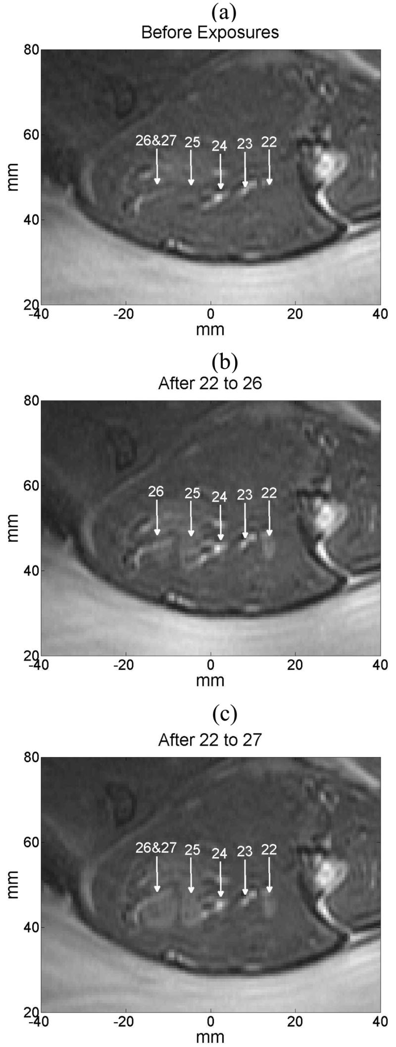
MR sagittal image where controlled exposures No. 22 to 27 (a) before any sonication was performed, (b) after exposures 22 to 26 were made, and (c) when exposure 27 was made (at the same location as exposure 26.

MR sagittal image where controlled exposures No. 22 to 27 (a) before any sonication was performed, (b) after exposures 22 to 26 were made, and (c) when exposure 27 was made (at the same location as exposure 26.