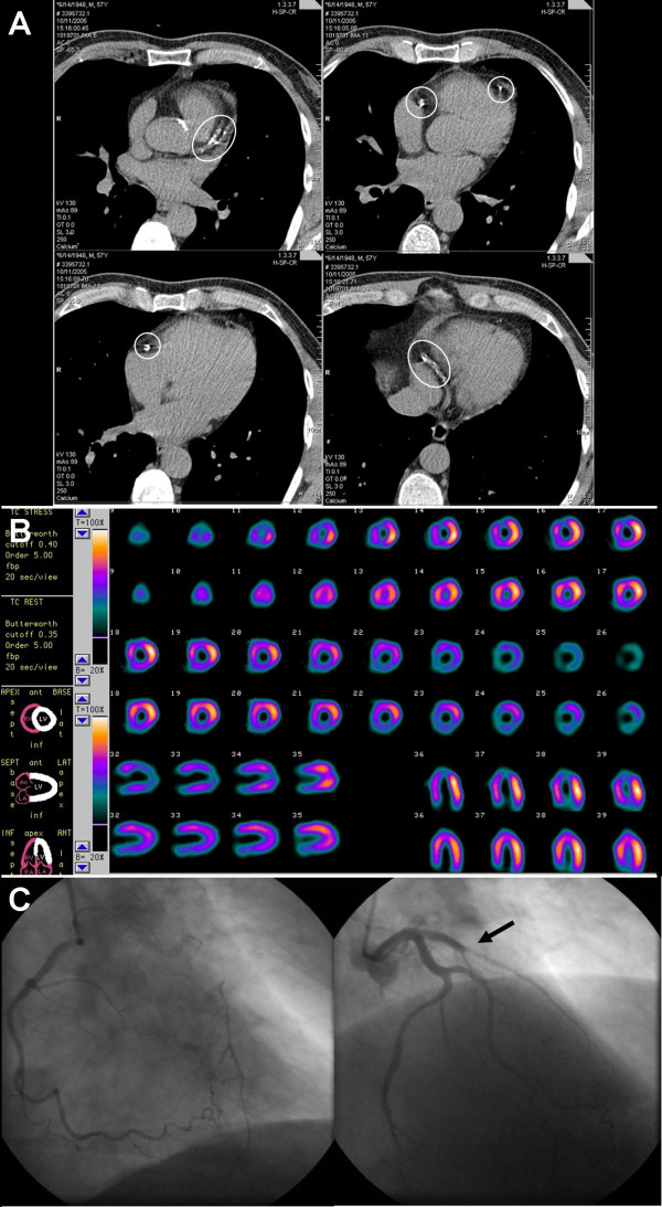
Figure 1.
Images of a 66-year-old symptomatic man who had a coronary artery calcification score of 671; A: electron-beam computed tomography, B: myocardial perfusion single photon emission computed tomography (SPECT), C: coronary angiography (CAG). Circles define regions of coronary calcification. SPECT demonstrated myocardial perfusion defect in the apex and anteroseptal region after bicycle stress test (upper rows). These were completely reversible during myocardial perfusion rest test. CAG revealed 1-vessel disease of the left anterior descending artery, with collateral filling from the right coronary artery. Arrow indicates the occlusion of the left anterior descending artery. Based on these results, the patient was accepted for coronary artery bypass graft surgery.

