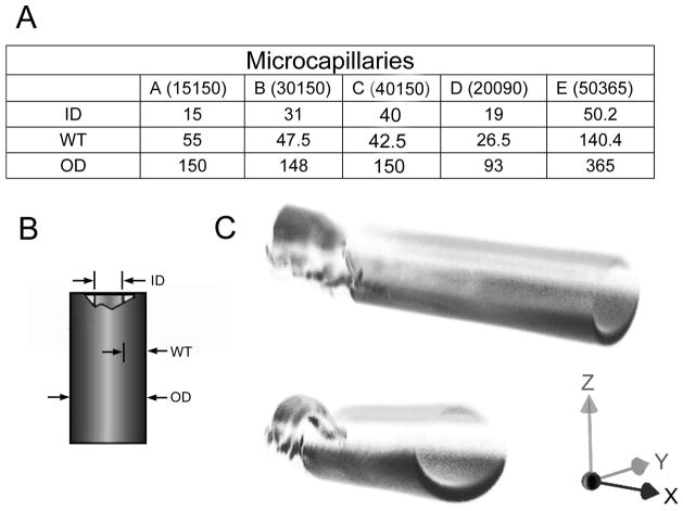
Figure 3.
The tissue phantoms were commercially available fused silica microcapillaries (Polymicro Technologies). 5 different sizes and wall thicknesses were chosen for this study (A). (B) The inner diameter (ID), wall thickness (WT) and outer diameter (OD) varied between capillaries (C) Structured illumination image of the capillaries with the polyimide coating resulted in partial imaging illustrated by the differential imaging of the fractured and unfractured portion of the capillary.

