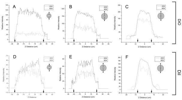
Figure 4.
Quantitative morphometry of the z-axis (optical axis) dimension of 3D reconstructed fused silica microcapillaries after structured illumination acquisition. Linescans from a DiO (green) and DiI (red) lipophilic dyes were acquired using 20x (solid line) and 40x (dotted line) magnification. No software compression was used.

