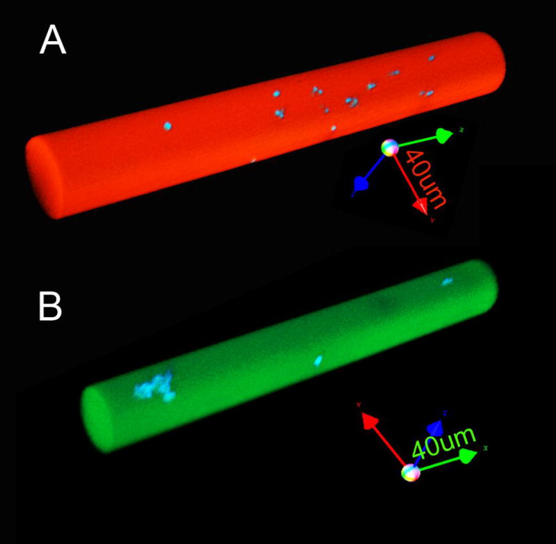
Figure 5.

3D reconstructions of fused silica capillaries after dual wavelength structured illumination acquisition. The capillaries were labeled with (A) DiI (red) and (B) DiO (green) lipophilic dyes containing microspheres labeled with coumarin (blue) derivatives. The images of the 50um diameter capillary were acquired using 20x objective without software compression (arrow length=40um).
