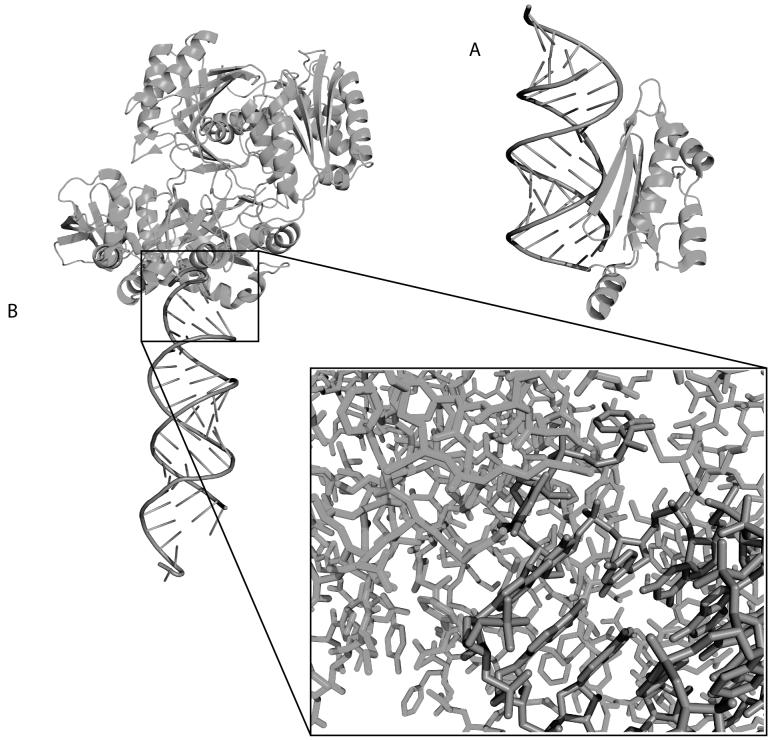
Figure 5.
Cartoon representations of the structures of RNA/Protein complexes important in RNA interefence. A) Crystal structure of the p19 protein viral suppressor bound to double-stranded RNA (PDB ID 1R9F). The alpha-helical arm binds to the bottom of the siRNA and is capped with Tryptophan residues that detect the exposed nucleotide bases at the ends of double-stranded RNAs. B) Crystal structure of the Argonaute protein bound to an siRNA (PDB ID 2F8S). The inset illustrates the complexity of the RNA/protein interface.

