Fig. 7.
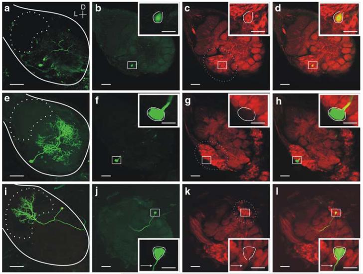
Immunohistochemistry revealed many olfactory neurons do not contain high levels of sGC-immunoreactivity. A subset of neurons in this study (Tables 1, 2) was labeled with lucifer yellow (LY) dye and subsequently tested for sGC-immunoreactivity (sGCir). a A whole-mount view of a LY filled Type Ib LN (LN28, see Fig. 5a) with a cell body in the lateral cell body cluster (dashed oval c) and no ramifications in the MGC (dashed oval in a). b The AL was frontally sectioned (100 μM sections) and c, labeled with the MsGCα1/Cy3 antibody (images show single optical sections 3 μM). A magnified view of the cell body is shown in the inset. d LN28 contained strong levels of sGCir. e-h, Another LN, Type Ia (LN17), was found to contain little or no sGCir. i-l sGC-immunohistochemistry in PN4 (Fig. 6d, e) revealed that while neighboring PNs appeared to contain strong labeling, this PN and its projection (arrow) did not contain high levels of sGCir. Calibration 100 μM; inset is 20 μM. Orientation D: dorsal; L: lateral

