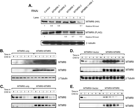
FIGURE 4.
Stability of MTMR6 and MTMR9 is increased by the formation of the complex. A, HeLa cells transfected with MTMR6-HA and MTMR9-FLAG for 24 h were treated with control or RNAi against MTMR6 (R6-1, R6-2), MTMR9 (R9-1, R9-2), or both (R6-1 + R9-1) for an additional 24 h. Cell lysates were analyzed by Western blotting for MTMR6 (HA) or MTMR9 (FLAG). β-tubulin levels are shown as a loading control. Relative R6 level, relative MTMR6 level; Relative R9 level, relative MTMR9 level. B, HeLa cells transfected with MTMR6-HA, or MTMR6-HA plus MTMR9-FLAG for 24 h. One hundred and fifty μg/ml cycloheximide (CHX) was added into the medium for the times indicated. The levels of MTMR6 and MTMR9 were detected by Western blotting with an anti-HA or anti-FLAG antibody, respectively. C, the same as B except with MTMR6 alone; the left six lanes contain 3-fold more total protein. D, HEK-293 cells stably transfected with MTMR9-FLAG were transiently transfected with MTMR6-HA. Six hours after the transfection, 0.5 μg/ml tetracycline was added into medium to induce the expression of MTMR9-FLAG. Eighteen hours later, cells were treated with cycloheximide for the indicated times. The levels of both MTMR9 and MTMR6 were detected by Western blotting with anti-FLAG or anti-HA antibody, respectively. Each sample contains equal amounts of total protein. E, the same as C except with MTMR9 alone; the left six lanes contain 3-fold more total protein.

