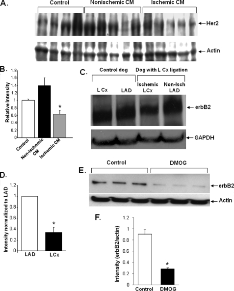
FIGURE 1.
Ischemia decreases the level of erbB2 protein in cardiomyocytes. A, Western blot with Her2 antibody on samples from normal human hearts (control) and from the hearts of patients with non-ischemic cardiomyopathy or ischemic cardiomyopathy. Four normal samples and five samples from ischemic and nonischemic cardiomyopathy patients were included in our studies. B, band densitometry was performed on the blot shown in A, and Her2 levels were normalized to actin levels (*, p = 0.011 versus control). C, Western blot for erbB2 protein levels in the hearts of dogs subjected to ischemia in the LCx artery for 2–5 h then immediately sacrificed. Tissue samples from the LCx (ischemic) and LAD (non-ischemic) territories were isolated along with similar samples from sham-instrumented (control) animals, which underwent all surgical procedures except coronary artery constriction, n = 3 in each group. D, quantification of erbB2 levels from Western blots similar to those shown in C. (*, p = 0.009 versus LAD). E, Western blot of NRCM treated with DMOG (a HIF-stabilizer) and control NRCM. F, quantification of erbB2 levels from the Western blots in E. DMOG treatment resulted in a significant decrease in the levels of erbB2 (*, p < 0.05 versus control). Band intensities were measured using ImageJ and normalized to the internal control (GAPDH or actin). Data are presented as mean ± S.E.

