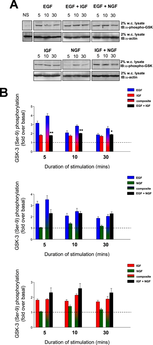
FIGURE 4.
Combined hormonal phosphorylation of GSK-3 in PC12 cells. A, representative Western blots depicting GSK-3 phosphorylation upon EGF, IGF, or NGF stimulation for 5, 10, and 30 min duration. Protein loading was controlled by assessing the presence of actin in each lane. The histograms in B depict means ± S.E. GSK-3 phosphorylation data from at least three independent experiments per histogram as demonstrated in A. EGF (blue bars), IGF (red bars), and NGF (green bars) indicate responses induced by individual ligand stimulation. Shaded mixed color bars indicate theoretical summation of individual responses compared with the actual resultant response induced by legend co-stimulation (black bars). IB, immunoblot; w.c. lysate, whole-cell lysate; NS, no stimulation.

