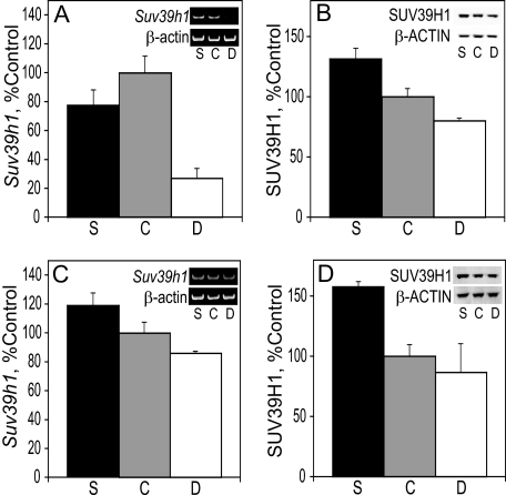
FIGURE 5.
Suv39h1 in the liver and cortex. A, RNA was isolated from the liver of E17 embryos and used for RT-PCR of Suv39h1 and normalized to β-actin (see example in gel insets). B, SUV39H1 protein levels in E17 liver were analyzed by immunoblot and normalized to β-ACTIN (see example in blot insets). C, RNA was isolated from the cortex of E17 embryos and used for RT-PCR of Suv39h1 and normalized to β-actin (see example in gel insets). D, SUV39H1 protein levels in E17 cortex were analyzed by immunoblot and normalized to the levels of β-ACTIN (see example in blot insets). See “Results” for statistics. Bar labels: S, choline-supplemented; C, controls; D, choline-deficient.

