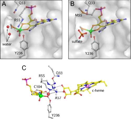
FIGURE 5.
Active site structures of SDHR55M and SDHH57A. A and B show a transparent surface at the substrate binding sites of SDHWT and SDHR55M, respectively. The water-filled cavity adjacent to the equatorial oxygen is larger in the mutated enzyme and is occupied by a sulfate ion. C, hydrogen bonding network around the active site of SDHH57A in a similar view as SDHWT in Fig. 1D. The two alternative positions for Arg-55 and the heme propionate group are shown. An additional water molecule occupies a site close to the pterin, and fulfills potential hydrogen bond contacts.

