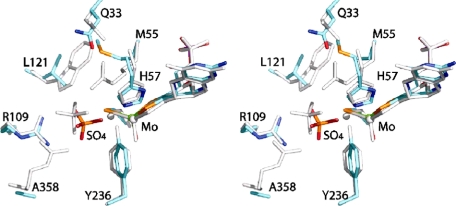
FIGURE 6.
Comparison of the position of bound sulfate in the active sites of SDHR55M and CSO. The stereoview shows the two active sites superimposed with the CSO residues colored gray and the the SDHR55M atoms colored as follows: molybdenum (green), sulfur (orange), phosphorous (magenta), oxygen (red), nitrogen (blue), and carbon (cyan). Labels show residues with SDH numbering.

