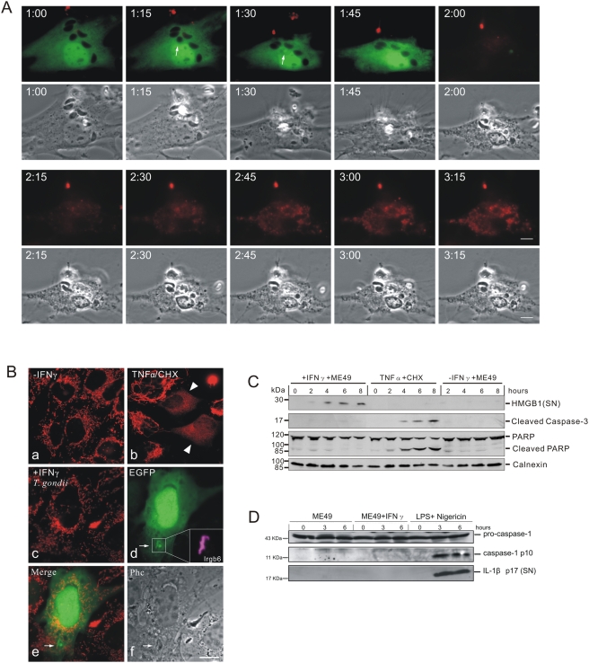
Figure 5. IFNγ-treated T. gondii infected host cells undergo necrosis after vacuolar disruption.
(A) Phosphatidylserine is not expressed on the cell surface before death. MEFs were induced with 200 U/ml IFNγ and transfected with pEGFP. After 24 hours they were infected with ME49 strain T. gondii for 1 hour at MOI between 5 and 10 and observed microscopically by time-lapse photography. Phosphatidylserine was detected by adding 1% (v/v) Alexa-555-labeled annexin V with 2.5 mM CaCl2 into the medium. Images were taken at 5 minute intervals and selected images from the series are shown. One intracellular T. gondii rounded up and was permeabilised at 1:30 (arrows) and the host cell died at 2:00. No annexin V signal was detected on the plasma membrane before cell death. After permeabilisation of the plasma membrane, annexin V accumulated steadily on intracellular material. Scale bar: 10 µm. The videos from which these frames were extracted are presented as Videos S11 and S12. (B) Cytochrome C is not released from mitochondria in cells containing a disrupted vacuole. MEFs were induced with 200 U/ml IFNγ and transfected with pEGFP for 24 hours (c–f). Cells were then infected with ME49 strain T. gondii at MOI between 5 and 10 for 4 hours, fixed and stained for cytochrome C (red) and Irgb6 (inbox in d). Arrows indicate the permeabilised T. gondii and Irgb6 staining shows the disrupted PVM. As control, MEFs without any treatment (a), or treated with TNFα (40 ng/ml) and cycloheximide (10 µg/ml) for 4 hours to induce apoptosis (b), were stained for cytochrome C. Arrowheads indicate cells showing release of mitochondrial cytochrome C. Scale bar 10 µm. (C) HMGB1 is released from IFNγ-induced cells infected with T. gondii. MEFs were treated with 200 U/ml IFNγ or left untreated for 24 hours and infected with ME49 T. gondii for the indicated times. Cells were then lysed and the lysates blotted for cleaved caspase-3 and PARP, as well as calnexin as loading control. Cell culture supernatants were collected at the indicated times after infection and blotted for HMGB1. MEFs treated with TNFα (40 ng/ml) and cycloheximide (10 µg/ml) were blotted as control for apoptotic cells. (D) Caspase-1 and IL-1β are not activated. BMMs were treated as described in (C). Cell lysates were blotted for caspase-1 and cell culture supernatant were blotted for IL-1β. BMMs were first treated with LPS (1 µg/µl) for 24 hours, and then treated with nigericin (20 µM) for the indicated times to activate inflammasomes as positive control.

