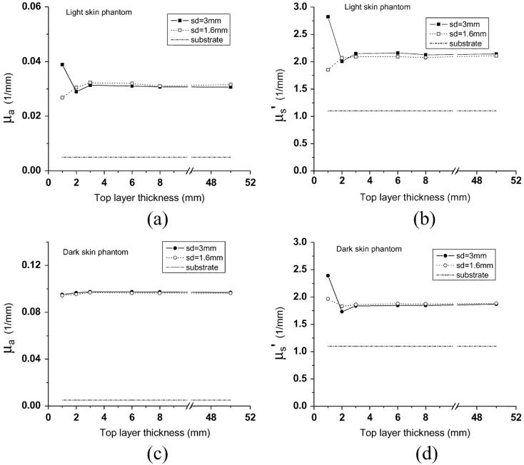
Fig. 2.
(a) μaand (b)μ′s recovered from light skin two-layer phantoms, and (c)μa and (d)μ′srecovered from dark skin two-layer phantoms. The thickness of the top layer of the two-layer phantoms varies from 1 to 8 mm. Two diffusing probes having source-detector separations of 3 mm (solid squares and solid circles) and 1.6 mm (squares and circles) were employed. Dash-dot lines represent optical properties of the substrate of the two-layer phantoms.

