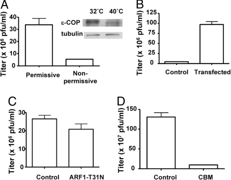
Fig. 2.
Characterizing how coatomer acts in vaccinia formation. (A) A CHO cell line that expressed a temperature-sensitive mutant of ε-COP was incubated either at permissive or nonpermissive temperature for 12 h, and then infected with a recombinant virus (vP30CP77, capable of replicating in CHO cells) for another 24 h. The vaccinia replication assay was then performed, with the mean and standard error from 3 experiments shown (P < 0.01). Gel shows the level of ε-COP, comparing permissive versus nonpermissive temperatures. (B) The mutant CHO cell line that expressed a temperature-sensitive mutant of ε-COP was stably transfected with wild-type ε-COP. After shifting to the nonpermissive temperature for 12 h followed by infection with the recombinant virus (vP30CP77) for 24 h, the vaccinia replication assay was then performed, with the mean and standard error from 3 experiments shown (P < 0.01). (C) HeLa cells, transiently transfected with ARF1-T31N for 48 h or mock-treated, were infected with WR virus for 24 h. The vaccinia replication assay was then performed, with the mean and standard error from 3 experiments shown (P = 0.15). (D) HeLa cells were infected with WR virus for 24 h, with CBM treatment (or mock treatment with vehicle alone) given at the start of the infection. The vaccinia replication assay was then performed, with the mean and standard error from 3 experiments shown (P < 0.01).

