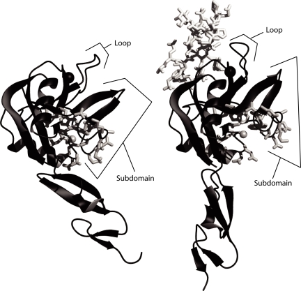
Fig. 1.
Opening of a cleft in the lectin domain in the extended selectin conformation. (Left) P-selectin in the unliganded conformation (PDB ID code 1G1Q). (Right) P-selectin in the liganded, extended conformation (1G1S). In each structure, the Cβ atom of Ala-28 in the cleft is shown as a gray sphere, and other side chains that line the cleft are shown as white sticks. The divalent cation in the ligand binding site is shown as a larger black sphere. Molecules are shown as ribbons in identical orientations after superposition on the lectin domain.

