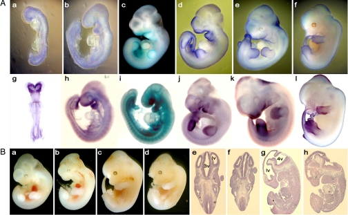
Fig. 3.
Morphological analysis of the head in mutant embryos and expression of Gtf2i and Gtf2ird1. (A) Expression of Gtf2i (a–f) and Gtf2ird1 (g–l) was detected by the β-galactosidase staining and whole-mount in situ hybridization of developing embryos. (B) Brain defects in Gtf2ird1 embryos at E11.5 (b) and E12.5 (d, f, and h) are compared with WT embryos (a, c, e, and g), respectively. (e–h) Parasagittal sections of WT and Gtf2ird1+/− embryos. (b, d, f, and h) Mutant brain shows bitemporal narrowing. lv, lateral ventricle; 3v, third ventricle; 4v, fourth ventricle.

