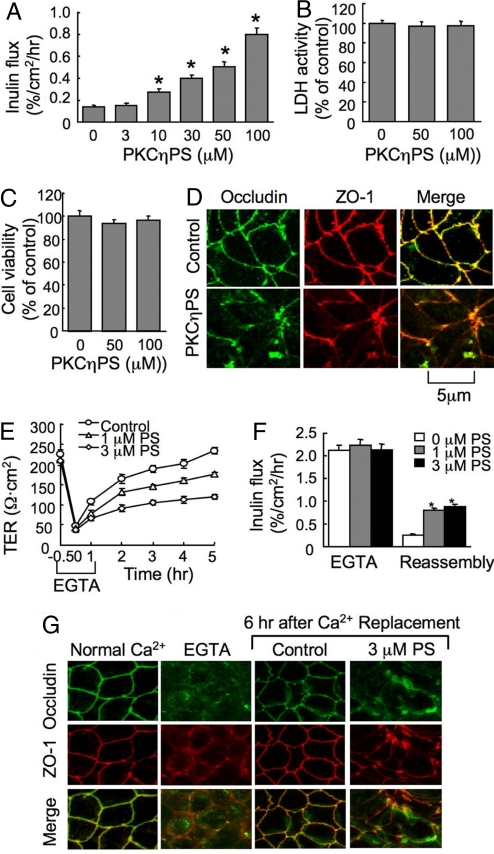
Fig. 1.
PKCηPS disrupts TJs in Caco-2 cell monolayers. (A) Cell monolayers were incubated with varying concentrations of PKCηPS. Inulin permeability was measured at 1 hour. (B and C) Cell monolayers were incubated with 50 or 100 μM PKCηPS for 2 hours, and the incubation medium was assayed for LDH activity (B) and the cells analyzed for cytotoxicity by WST assay (C). (D) After 1-hour incubation with 50 μM PKCηPS, cell monolayers were fixed and double labeled for occludin and ZO-1. (E and F) Cell monolayers were incubated with EGTA to deplete calcium. TJ reassembly was induced by calcium replacement in the absence or presence of PKCηPS (PS). TJ assembly was evaluated by measuring TER (E) and inulin permeability (F). (G) At different stages of calcium switch-mediated TJ assembly with or without PKCηPS (PS), cell monolayers were double labeled for occludin and ZO-1 by immunofluorescence method. Values in A–C, E, and F are mean ± SEM (n = 6), and the asterisks indicate the values that are significantly different from corresponding values for cells incubated without PKCηPS.

