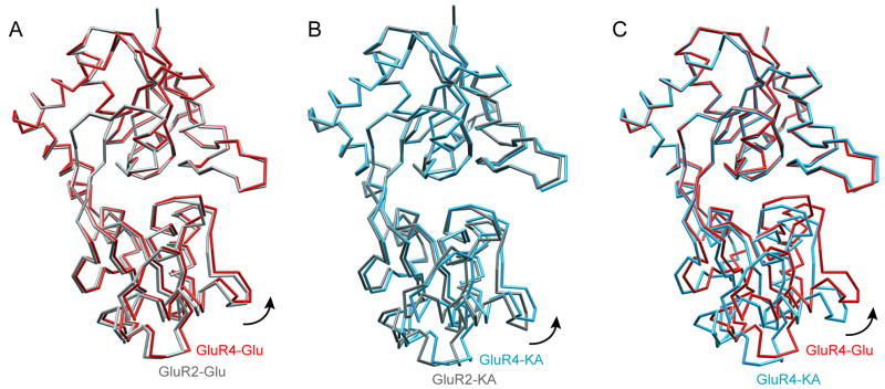
Figure 2. Comparison of cleft closure in GluR4 and GluR2 ligand-bound structures.
(A) Superposition of glutamate-bound GluR4 LBD (red) and GluR2 LBD (gray) structures. (B) Superposition of the kainate-bound GluR4 LBD (blue) and GluR2 LBD (gray) structures. (C) Superposition of GluR4 LBD structures bound to glutamate (red) and kainate (blue). The Glu-bound structure shows a greater degree of cleft closure than the kainate-bound structure (difference ~6.7°). In each case, the core residues of lobe 1 were superimposed. This figure was prepared using CHIMERA (36)

