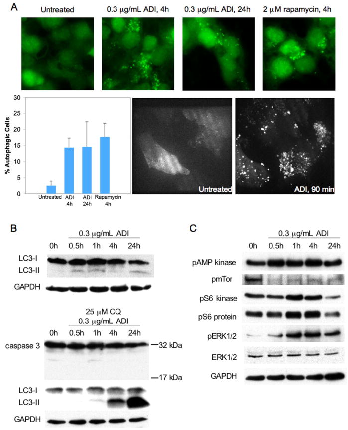
Figure 4. ADI-PEG20 induces autophagy in CWR22Rv1.
A, CWR22Rv1 cells overexpressing eGFP-LC3 were treated with 0.3μg/mL ADI-PEG20 for 1.5, 4, and 24 hours, or 2 μM rapamycin for 4 hours. Punctae represent autophagosome formation. Autophagic cells were quantified from random image fields totaling 200 cells and reported as mean±SE. B, Immunoblot for 0.3μg/mL ADI-PEG20 timecourse of CWR22Rv1. α-LC3 detects LC3-I and LC3-II. Autophagic flux was confirmed by co-administering 25μM chloroquine with 0.3μg/mL ADI-PEG20. α-Caspase-3 detects the 32kDa pro-form and the activated 17kDa cleavage product. C, Immunoblot for 0.3μg/mL ADI-PEG20 timecourse of CWR22Rv1 using α-phopsho-AMP kinase, α-phospho-mTor, α-phospho-S6 kinase, α-phospho-S6 protein, α-phospho-ERK1/2, and α-ERK1/2. Loading control for phospho-mTor was verified using α-tubulin (not shown).

