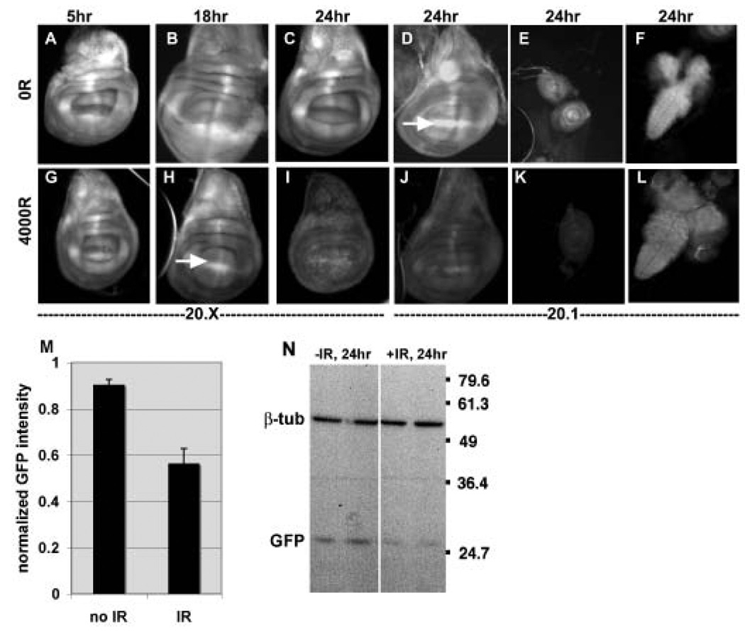
FIGURE 2. bantam sensor expression is reduced in irradiated imaginal discs.
Wing imaginal discs (A–D and G–J), leg imaginal discs (e and K) and the CNS (brains and Ventral Nerve Cord; F and L) from 3rd instar larvae were imaged for EGFP fluorescence at 5, 18 or 24 hr after exposure to 0 or 4000R of X-rays. Each irradiated tissue was imaged, and the images processed identically as for un-irradiated counterparts to allow direct comparison (e.g. unirradiated wing discs to irradiated wing discs; unirradiated leg discs to irradiated leg discs). ‘20.1’ and ‘20.X’ refer to different insertion lines that carry the Tub-EGFP sensor (Brennecke et al., 2003). Note that local enrichment of ban sensor expression, for example at the D/V boundary (Brennecke et al., 2003), is visible in some discs (arrows in D and H) but not others because of the focal plane shown.
(M) Mean GFP fluorescence from larval wing imaginal discs 24 hr after exposure to 0R (no IR) or 4000–5000R of X-rays (IR) were normalized and averaged as explained in Methods. The data are from 15 (no IR) and 15 (IR) discs in 4 different experiments of both 20.X and 20.1 insertion lines. Because of sample thickness, images from more than one focal plane were considered in determination of mean intensity. The decrease in GFP signal in irradiated discs is significant (p=0.0017). Error bars=1 SEM.
(N) The level of EGFP was analyzed by Western blotting of extracts from CNS and imaginal discs from larvae 24 hr after exposure to 0R (−IR) or 4000R (+IR) of X-rays. Duplicate samples are blotted for each condition. Extracts from equivalent number of larvae were loaded and β-tubulin serves as a loading control.

