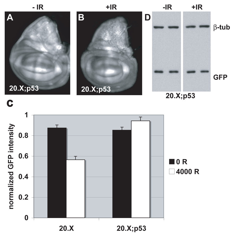
FIGURE 4. ban sensor expression remains unchanged after irradiation in p53 mutants.
(A, B) Wing imaginal discs from control (A) and irradiated larvae (B) are imaged for EGFP fluorescence 24 hr after irradiation with 4000 R of X-rays. Larvae were homozygous for p535A-1-4 and the 20.X ban sensor.
(C) Mean GFP fluoresence intensity for wing imaginal discs analyzed as described in Fig. 2M, at 24 hr after irradiation with doses shown. The decrease in GFP signal in irradiated discs is significant for the controls (20.X) (p=0.00004) but not for p53 mutants (20.X; p53) (p=0.14). The data are from 14 (20.X, no IR) and 10 (20.X, IR), 14 (20.X; p53, no IR) and 12 (20.X; p53, IR) discs in 2 different experiments. Error bars=1 SEM. 20.X = homozygous ban sensor. 20.X; p53 = the same in homozygous p535A-1-4 background.
(D) The level of EGFP in the above experiment was analyzed by Western blotting of extracts from larval CNS and imaginal discs from homozygous p535A-1-4 larvae that carry the 20.X ban sensor. No change in GFP was detected. Duplicate samples were blotted for each condition. Extracts from equivalent number of larvae were loaded and β-tubulin serves as a loading control.

