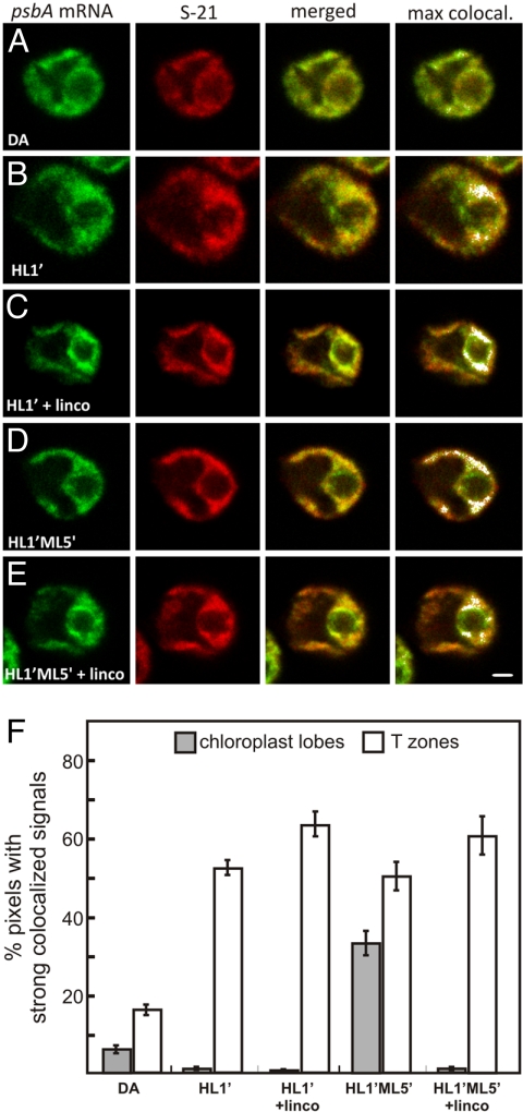
Fig. 3.
Localization of the psbA mRNA for the de novo assembly and repair of PSII. (A–E) Fluorescence signals from the psbA mRNA and the chloroplast ribosomal protein S-21 in cells from the following conditions: a 2-h DA (A); DA cells exposed to HL for 1 min (HL1′) to induce psbA translation for de novo PSII assembly (B); HL1′ cells generated in the presence of lincomycin (C); HL1′ cells exposed to ML for 5 min, a condition of PSII repair (HL1′ML5′) (D); and HL1′ML5′ cells generated in the presence of lincomycin (E). The micrographs show 0.2-μm optical sections. (Scale bar: 1 μm.) (F) The percentages of pixels with strong colocalized signals in T zones (white bars) and chloroplast lobes (shaded bars) across all cells from the 5 conditions. The error bars indicate 2 standard errors. For each experiment, n ≥ 20 cells.

