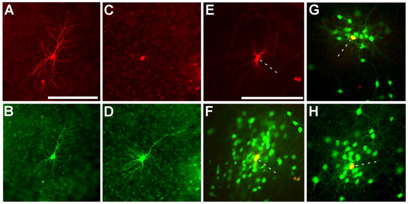
Figure 2. Selective Infection and In Situ Complementation in Slice Culture.
(A–D) Initial infection is restricted to cells expressing the ASLV-A receptor, TVA. In these control experiments to test infection selectivity, isolated neurons in cultured brain slices were transfected using the gene gun with two genes, one encoding TVA, and the other, DsRed2. ASLV-A-pseudotyped rabies virus [SADΔG-EGFP(EnvA)] was applied the next day and images were taken 6 days following. (A and C) DsRed2 expression marking transfection with TVA and (B and D) EGFP expression indicating subsequent selective infection of the same TVA-expressing cells with pseudotyped virus. (E–H) In situ complementation permits transsynaptic spread from a single initially infected cell to a cluster of monosynaptically connected cells. Isolated neurons were transfected with three genes encoding TVA to permit viral infection, DsRed2 to mark transfected cells, and the rabies virus glycoprotein gene to complement the deletion in the viral genome. (E) DsRed2 fluorescence indicating a transfected cell, marked with a dotted line, at the center of the cluster shown in (F). (G and H) Two more examples of clusters surrounding a single transfected cell. The initially infected cells expressing both EGFP and DsRed2 appear yellow. Scale bars: 200 μm.

