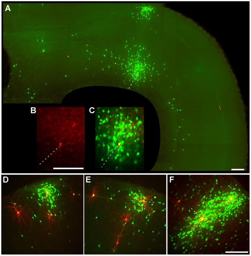
Figure 3. More Examples of Transcomplemented Tracing.
(A–C) Long-range viral spread from a single initially infected cell. (A) A huge cluster of green cells surrounds a single red/green deep-layer cortical neuron (dotted line) at 8 days postinfection. Another dense cluster of cells is also infected in the superficial cortical layers immediately above it, consistent with known projections of superficial layers to deeper ones; distant deep-layer pyramidal cells are also infected, again consistent with known patterns of long-range intralaminar connectivity. To the left of the putatively initially infected cell is a second yellow (double-labeled) cell, apparently secondarily—and recently—infected because of the lack of green cells surrounding it. (B–C) Closeup of central cluster from (A). (D–F) More examples of in situ complementation: clusters of infected cells surrounding isolated putatively postsynaptic ones. Scale bars: 200 μm.

