
Figure 1.
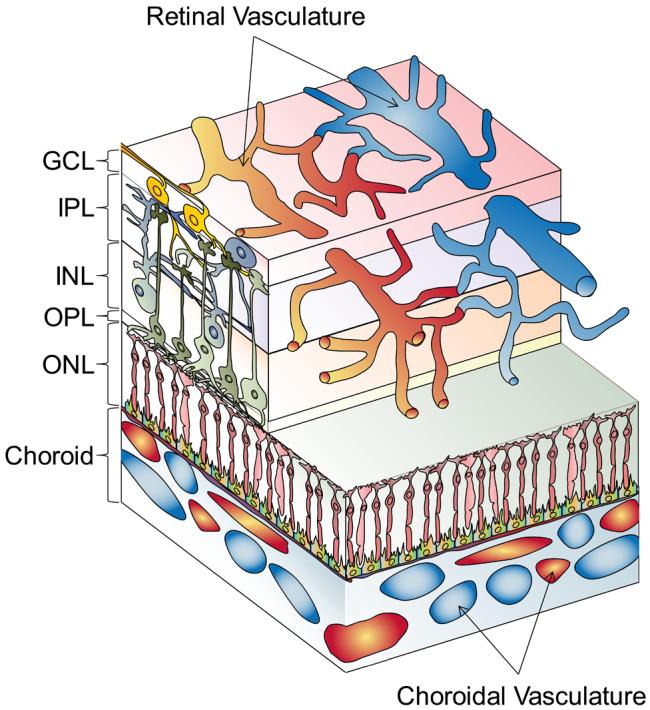
Diagram of the neural retina and its vascular supplies (not to scale). The layers of the neural retina (ganglion cell, inner plexiform, inner nuclear, outer plexiform, outer nuclear) are indicated. Blood flow through the choroidal vessels is swift. The retinal vasculature, visible by ophthalmosocopy, lies among the ganglion cells on the vitreal surface of the retina and extends capillary networks deep into the postreceptor layers. The caliber of the retinal arterioles adjusts to perturbations in blood oxygen levels (“autoregulation”).