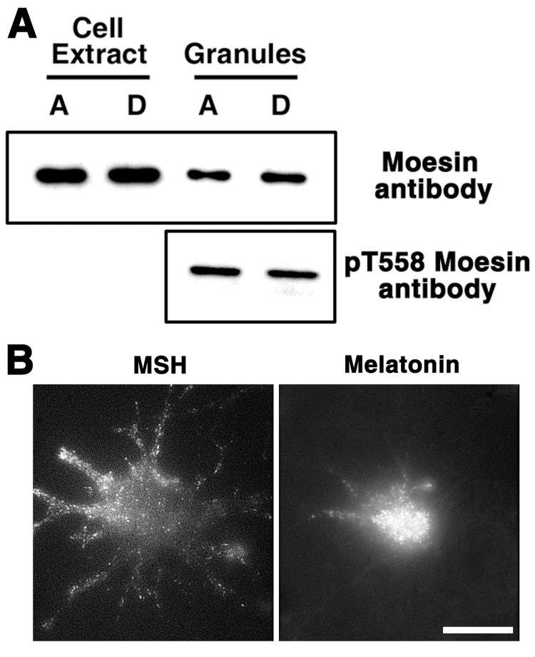
Figure 3. Moesin binds pigment granules in vivo and in vitro.
A, Top, immunoblotting with anti-moesin antibody of cell extracts or pigment granules prepared from melanophores with aggregated (A) or dispersed (D) pigment granules; bottom, immunoblotting with pT558 antibody, which recognizes the activated form of moesin, of pigment granule preparations isolated from cells with aggregated or dispersed pigment granules. Moesin in the activated form is bound to pigment granules. B, Fluorescence images of the same cell transfected with moesin-GFP treated with MSH or melatonin to induce aggregation or dispersion of pigment granules. Moesin is localized to fluorescent dots whose distribution and behavior in response to hormones is similar to pigment granules. The scale bar is 20 μm.

