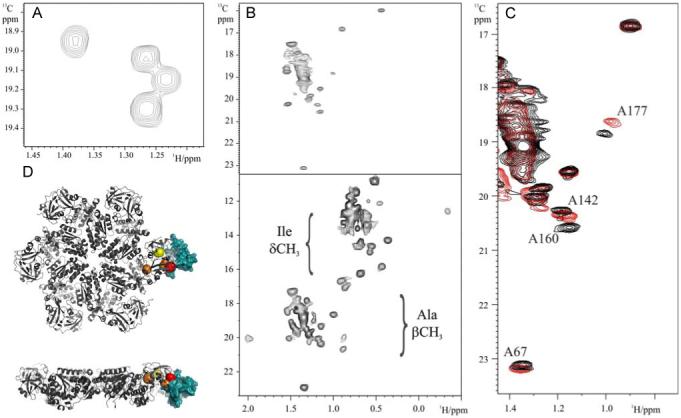
Figure 1.

Alanine labeling and 1H-13C 2D Methyl TROSY spectra of (A) 13CH3-Ala,U-2H labeled Ufd1in UN (B) 13CH3-Ala,U-2H labeled p97 ND1 (top) and 13CδH3-Ile, 13CH3-Ala,U-2H labeled p97ND1 (bottom) (C) Expanded region of the 1H-13C 2D Methyl TROSY spectrum of 13CH3-Ala,U-2H labeled p97 ND1 in the absence and presence of perdeuterated Npl4 UBD. (D) Structural model of the p97 ND1-Npl4 UBD complex. The p97 hexamer is shown as a grey ribbon and perturbed alanine residues are indicated by red (most shifted), orange (intermediate) and yellow (least) balls. The position of Npl4 UBD (blue surface) in complex with p97 N domain is shown for one of monomers. Details are given in supplementary information.
