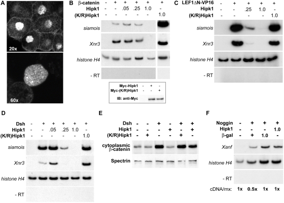
Figure 4. Hipk1 is nuclear and acts at the level of transcription during X. laevis development.
(A) Confocal micrographs of animal pole explants recombinantly expressing Myc-tagged Hipk1 and visualized with anti-Myc antibody. Hipk1 localizes strongly to the nucleus and nuclear speckles, with low-levels of apparent extra-nuclear signal near the plasma membrane. (B–D) Hipk1 over-expression blocks activation of the Wnt-responsive genes siamois and Xnr3 downstream of β-catenin (B), Lef/Tcf (C), and Dsh (D). X. laevis embryos were uninjected (lanes furthest on left in all panels) or injected with synthetic RNAs encoding β-catenin (B), LEF1ΔN-VP16 (C) or Dsh (D). Nanogram quantities of RNAs encoding Myc-Hipk1, Myc-(K/R)Hipk1 or β-galactosidase were co-injected as indicated. RT-PCR was used to assess target gene activation; histone H4 was assessed as a loading control, and reactions without reverse transcriptase (−RT) performed to rule out genomic DNA contamination. (B, inset) Myc-tagged Hipk1 and (K/R)Hipk1, injected at 1 ng per blastomere, are expressed at comparable levels in X. laevis as demonstrated by western blot. (E) Over-expression of Dsh in HEK293T cells leads to an increase in cytoplasmic β-catenin that is not affected either positively or negatively by over-expression of Hipk1. (F) Induction of Xanf by Noggin in X. laevis animal caps is not affected by over-expression of Hipk1.

