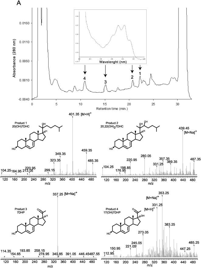
Figure 4. Metabolism of 7DHC by pig adrenal glands.
Fragments of pig adrenal glands were incubated with 0.5 mM 7DHC. Steroids were extracted with methylene chloride and subjected to HPLC and UV spectral analyses. The UV spectrum for all peaks is the same (see arrows). The mass spectra of the collected peaks 1–4 were obtained by direct injection into the MS module of the Bruker Esquire-LC/MS Spectrometer.

