
Fig. 2.
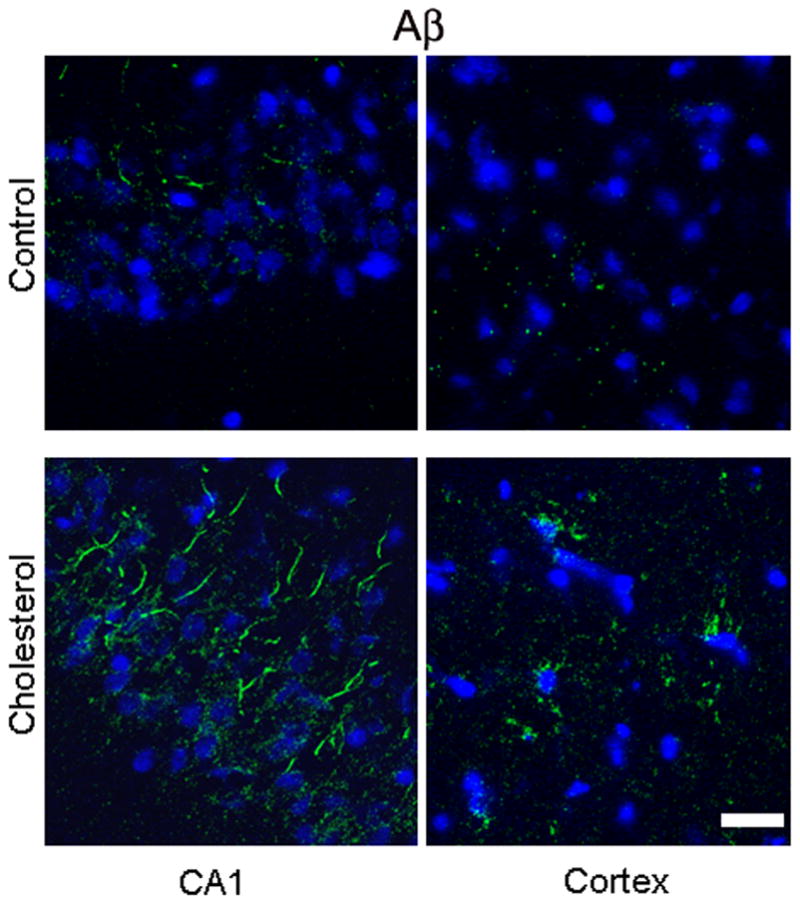
Immunofluorescence staining showing increased expression of Aβ levels (green) as detected by 6E10 antibody in the CA1 region of the hippocampus and in the adjacent cortex of a cholesterol-fed rabbit brain compared to a control rabbit. DAPI (blue) was used to stain nuclei. Bar= 20μm.