
Fig. 4.
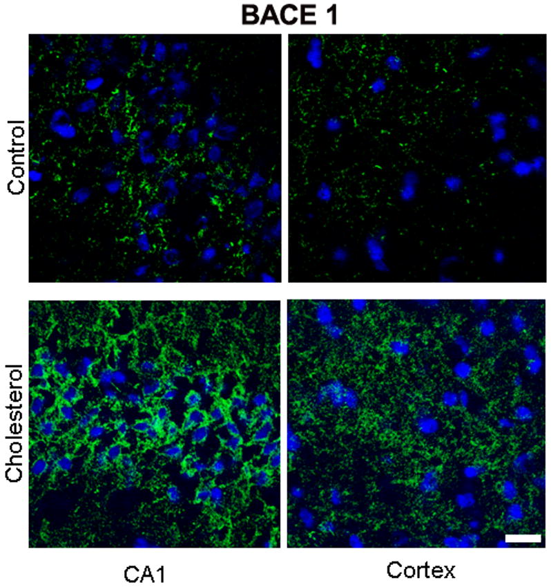

(A) Immunofluorescence staining demonstrating a dramatic increase in expression of BACE1 (green) in the CA1 region of the hippocampus and adjacent cortex in sections from a cholesterol-fed rabbit brain compared to section from a control rabbit. DAPI (blue) was used as nuclear counterstain. Bar= 20μm.
(B) Immunostaining for RAGE is low in sections from CA1 region of hippocampus or adjacent cortex in a control rabbit. In section from CA1 and adjacent cortex from a cholesterol-fed rabbit, immunoreactivity to RAGE (green) is increased in hippocampus and in cortex. Nuclei were stained with DAPI (blue). Bar= 20μm