
Fig. 6.
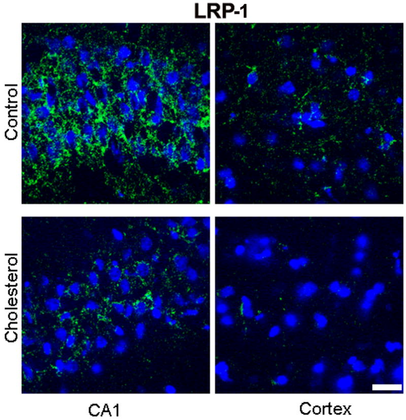
Immunofluorescence staining for LRP-1 (green) showing a marked immunoreactivity in the CA1 region of the hippocampus and a lesser reactivity in cortex from a control rabbit. In a cholesterol-fed rabbit, the immunoreactivity to LRP-1 antibody is greatly reduced in both hippocampus and cortex. DAPI (blue) was used as nuclear marker. Bar=20μm.