
Fig. 7.
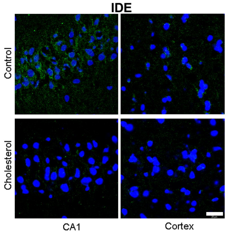
Immunofluorescence staining for IDE (green) is higher in the CA1 region of the hippocampus than the cortex of a control rabbit. IDE immunostaining is greatly reduced in both hippocampus and cortex from a cholesterol-fed rabbit. DAPI (blue) was used as nuclear marker. Bar=20μm.