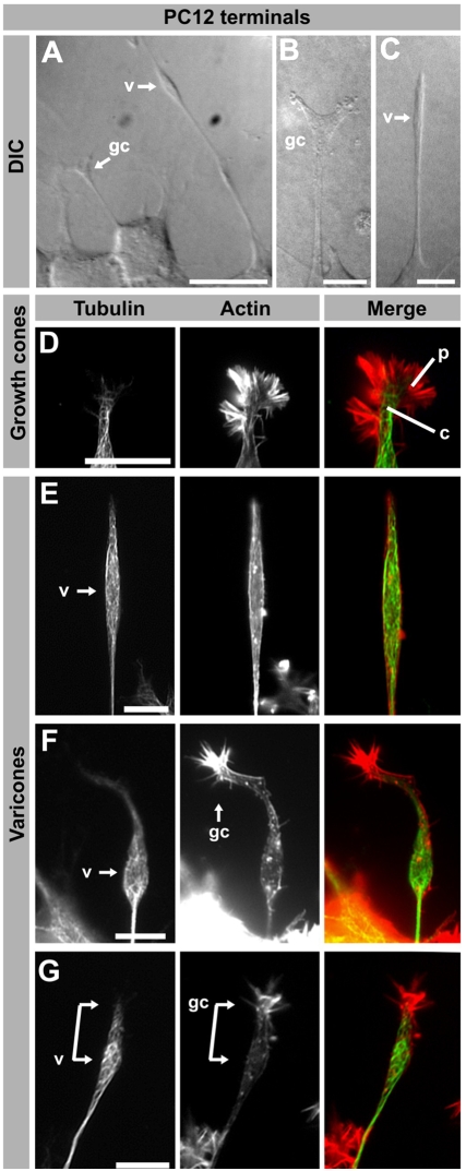
Figure 1. Morphological characterization of PC12 varicones and growth cones.
(A–C) Examples of differentiated PC12 cells observed using Differential Interference Contrast (DIC). A large varicosity at the tip of many neurites is apparent (“v” in A and C) while other terminals resemble neuronal growth cones (“gc” in A and B). (D–G) Fluorescence images of several PC12 cell neurite terminals labeled with a β-III-tubulin antibody and phalloidin to visualize tubulin and actin. While some terminals display a morphology similar to neuronal growth cones, including a tubulin-rich central domain (c) surrounded by an actin-rich peripheral domain (p) as shown in D, others have a large varicosity (E–G) and “growth cone” that can be collapsed (E) or more clearly visible (F–G). In some neurites, both the varicosity (v) and the growth cone (gc) appear as a connected structure (connected arrows in E). Scale bar A = 20 µm, B–G = 10 µm.

