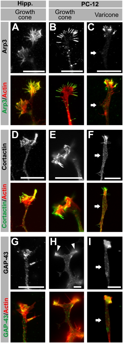
Figure 2. Immunocytochemical characterization of PC12 terminals I: growth cone markers.
(A–I) Fluorescence images of hippocampal neuron growth cones, PC12 growth cones and PC12 varicones, immunolabeled with growth cone markers (white or green) and co-labeled with phalloidin (actin, red). Arp3 (A–C), Cortactin (D–F) and GAP-43 (G–I) localize to the actin-rich regions of the growth cones, including the actin-rich region associated with varicones (C, F, I), but not to the varicosity associated with varicones (white arrow). Scale bar = 10 µm.

