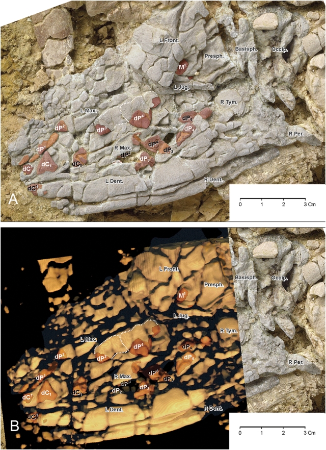
Figure 8. Fetal skull of Maiacetus inuus (GSP-UM 3475b).
(A)–Photograph showing bones (shaded white) and teeth (shaded brown) in lateral view. (B)–CT image overlain on tooth-bearing part of fetal skull in lateral veiw. Abbreviations: left frontal (L. Front.), left jugal (L Jug.), occipital (Occip.) with left and right condyles flanking the foramen magnum, and left and right dentaries (L dent., R dent.). Teeth are shaded brown, with darker brown representing enamel, and lighter brown representing roots and exposed dentine. White and black labels identify teeth of the left and right sides, respectively. Dotted lines trace outlines of fully formed crowns of left dP3 and dP4. These crowns are visible on the surface (A) where thin bone of the maxilla is pressed over more rigid underlying crowns, and as denser masses in the CT scan (B). Remaining teeth are identified by size and position relative to dP3 and dP4. Note presence of the developing crown of permanent left M1 posterodorsal to the crown of left dP4 (dorsal to the left jugal and posterior to the left frontal). Partial crown of right M1 (unlabeled) is visible just below and posterior to left M1.

