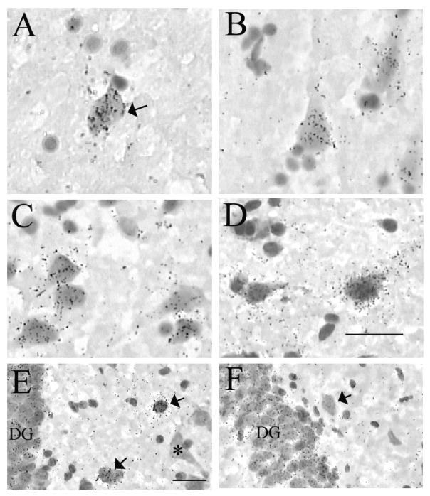
Figure 3.
Cellular expression of (A-C, E) NTNG1 and (D, F) NTNG2 mRNAs in (A-D) human temporal lobe and (E, F) P42 rat hippocampal formation. A: NTNG1 mRNA is expressed by occasional putative hippocampal interneurons (arrow), as observed here in the stratum oriens of CA3. B: NTNG1 mRNA over pyramidal neurons in the subiculum. C: NTNG1 mRNA expression by layer 3 perirhinal cortical neurons. D: NTNG2 mRNA in the perirhinal cortex is localized over deep layer 5/6 neurons. E: NTNG1 mRNA is expressed by granule cells of the rat DG, and by putative interneurons (arrows), but is not reliably detected over pyramidal neurons (asterisk). F: NTNG2 mRNA is expressed by granule cells of the rat DG, but is not reliably detected over putative hippocampal interneurons (arrow). Bars in (D) and (E) = 30 μm. Abbreviations as in Figure 2.

