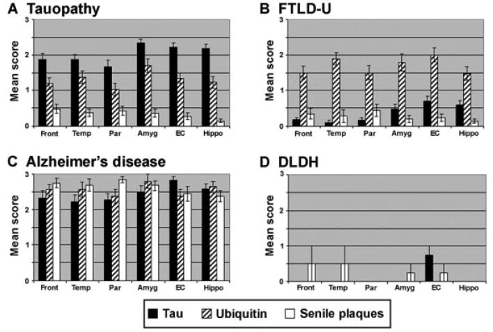
Fig 3.

Topographical distribution of pathological inclusions in frontotemporal dementia (FTD) subgroups. Semiquantitative distribution of tau (black bars), ubiquitin (striped bars), and senile plaque (white bars) inclusion pathology in FTD subgroups. The tau pathology in the Alzheimer’s disease (AD) patients was largely Thioflavine S–positive, whereas in the tauopathies, the pathology was largely Thioflavine S–negative. This is reflected in the Braak stage for each of the subgroups: (A) tau = 1.7 ± 1.1; (B) frontotemporal lobar degeneration with ubiquitin-positive inclusions (FTLD-U) = 0.8 ± 0.7; (C) AD = 5.8 ± 0.8; (D) dementia lacking distinctive histopathology (DLDH) = 0.8 ± 0.5. Amyg = amygdala; EC = entorhinal cortex; Front = middle frontal gyrus; Hippo = hippocampus, CA1/subiculum; Par = inferior parietal lobule; Temp = superior temporal gyrus.
