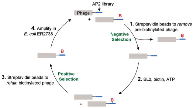
Figure 2.
Phage display selection scheme. The AP2 library (blue) is fused to the pIII coat protein. B represents biotin. After phage production in E. coli strain ER2738, a fraction of the phage pool is pre-biotinylated by endogenous BirA. In step 1 of the selection, streptavidin-coated beads are used to remove these pre-biotinylated phages (negative selection). In step 2, the remaining phage are treated with the BL2 enzyme. In step 3, streptavidin-coated beads are used to isolate the biotinylated phage from this mixture (positive selection). Finally (step 4), the recovered biotinylated phage are amplified in ER2738 for another round of selection.

