Figure 1.
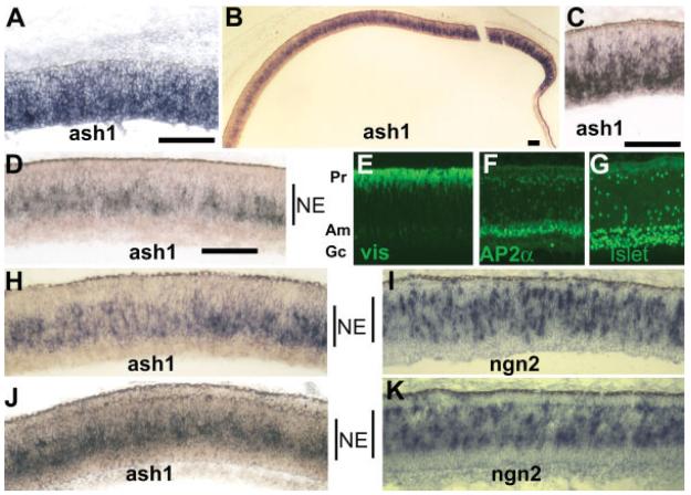
Expression of ash1 in the developing chick retina detected with dig-labeled antisense RNA probe. A: ash1 expression in E6 retina. B: ash1 expression at E8. C: Higher magnification of a peripheral region at E8. D: Higher magnification of a central region at E8. E–G: Immunostaining to mark the anatomical locations in a pseudostratified E8 retina of photoreceptor cells (Pr, visinin+, E), amacrine cells (Am, AP2α+, F), and ganglion cells (Gc, Islet-1+, G). H–K: Comparison of the spatial distribution of cells expressing ash1 (H, J) with that of cells expressing ngn2 (I, K), in developmental stage-matched regions of E8 retinas (H, I; and J, K). The neuroepithelial zone (NE) within the developing retina is marked, approximately, by a vertical line. Scale bars: 100 μm.

