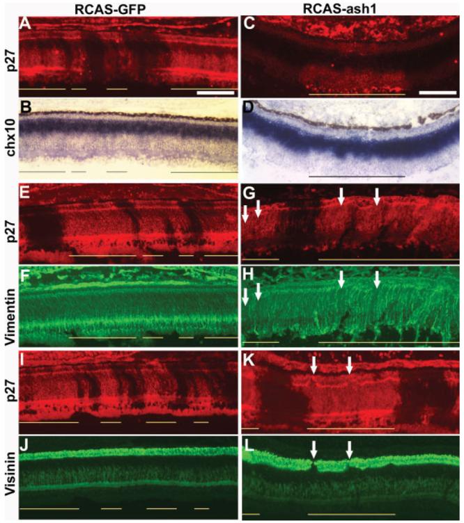
Figure 4.

Reductions in retinal cell populations in retinas infected with RCAS-ash1. A, B: Double-labeling of an E10 control retina partially infected with RCAS-GFP for p27 to identify viral infection (A) and for chx10 expression (in situ hybridization) to identify bipolar cells (B). C, D: Double-labeling for viral protein p27 (C) and for chx10 mRNA (D) of an E10 retina partially infected with RCAS-ash1. E–H: Double-labeling for viral protein p27 (E, G) and for vimentin (F, H) of E10 retinas infected with RCAS-GFP (E, F) or RCAS-ash1 (G, H). Arrows point to regions missing vimentin+ cellular processes. I–L: Double-labeling for viral protein p27 (I, K) and for visinin (J, L) of E10 retinas infected with RCAS-GFP (I, J) or RCAS-ash1 (K, L). Arrows point to regions lacking visinin+ cells. Scale bars: 100 μm.
